
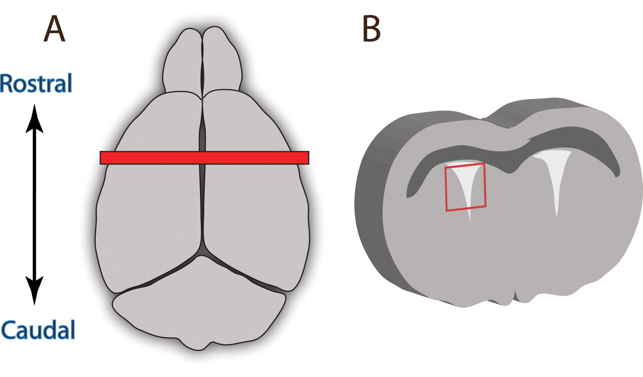
Figure 2.
(A) Dorsal view of the adult mouse brain. The red bar indicates the location of the cut used to isolate periventricular tissue. (B) The resulting cross-section of tissue with lateral ventricles exposed. Periventricular tissue should be dissected where indicated by the red box.


