
Cover image
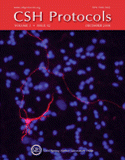
Cover Illustration: Fluorescence image of a GFP-transfected cerebellar granule neuron overlaid with Hoescht staining to identify nuclei of all cells in culture. The cells were transfected on DIV1 and fixed on DIV5, then subject to immunostaining for GFP (labeled with a Cy3-conjugated secondary). (Image courtesy of Parizad M. Bilimoria and Azad Bonni.) For more information on this culture technique, see Cultures of Cerebellar Granule Neurons (doi:10.1101/pdb.prot5107).

