
Cover image
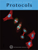
Cover Illustration: Composite fluorescence image of cultured HeLa cells at different stages of the cell cycle. Cells are expressing a recombinant mannosidase II-EGFP targeted to the Golgi apparatus (green), then immunolabeled for tubulin (red) and counterstained for DNA (blue). Multiphoton microscopy using a BioRad RTS2000-MP imaging system on a Nikon TE300 inverted microscope and a Nikon 60X 1.45 objective lens. (Image courtesy of Thomas Deerinck and Mark Ellisman.) For more information on fluorescent proteins and their variants, see Fluorescent Protein Tracking and Detection: Fluorescent Protein Structure and Color Variants (doi:10.1101/pdb.top63).

