
Cover image
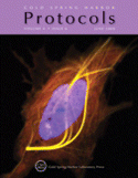
Cover Illustration: Fluorescence image of a zebrafish fibroblast (PAC2) co-transfected with H2BCFP, which labels the nucleus in blue, and DCXCitrine, which labels microtubules in yellow. Pac2 cells were cultured and transfected in imaging chambers. Image was taken 24 h after transfection with a 63X oil objective using a Zeiss LSM510. (Image courtesy of Martin Distel.) For more information on this method, see Culturing and Transfecting PAC2 Zebrafish Fibroblast Cells (doi:10.1101/pdb.prot5235).

