
Time-lapse video of primordium migration in zebrafish
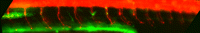
The primordium and neuromasts fluoresce in green owing to a cldnb:gfp insertion (Gilmour D, Knaut H, Maischein HM, Nusslein-Volhard C. 2004. Towing of sensory axons by their migrating target cells in vivo. Nat Neurosci 7: 491-492) and the sensory neurons fluoresce in red owing to a nbt:dsred insertion (Peri F, Nüsslein-Volhard C. 2008. Live imaging of neuronal degradation by microglia reveals a role for v0-ATPase a1 in phagosomal fusion in vivo. Cell 133: 916-927). Afferent axons accompany the primordium all along its migration (Metcalfe WK, Kimmel CB, Schabtach E. 1985. Anatomy of the posterior lateral line system in young larvae of the zebrafish. J Comp Neurol 233: 377-389; Gilmour D, Knaut H, Maischein HM, Nusslein-Volhard C. 2004. Towing of sensory axons by their migrating target cells in vivo. Nat Neurosci 7: 491-492). The upper red stripe is the spinal cord; the oblique fibers are motor axons. [Supplemental movie from Imaging in Developmental Biology: A Laboratory Manual, Cold Spring Harbor Laboratory Press.]


