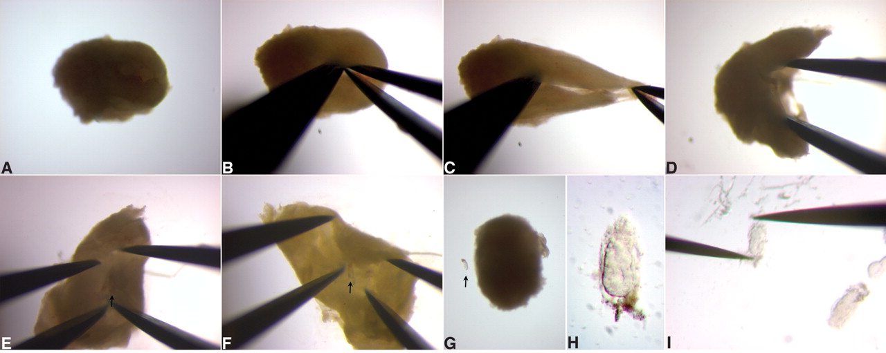
Figure 2.
(A-F) Sequence of steps for isolating an E5.5 embryo from a deciduum. (G) An embryo and deciduum next to each other to provide a sense of scale. The embryo in this panel is ~100 μm in width. The arrow in E-G points to the embryo. (H) An isolated E5.5 embryo, with intact Reichert’s membrane. (I) Removal of the Reichert’s membrane with tungsten needles.