
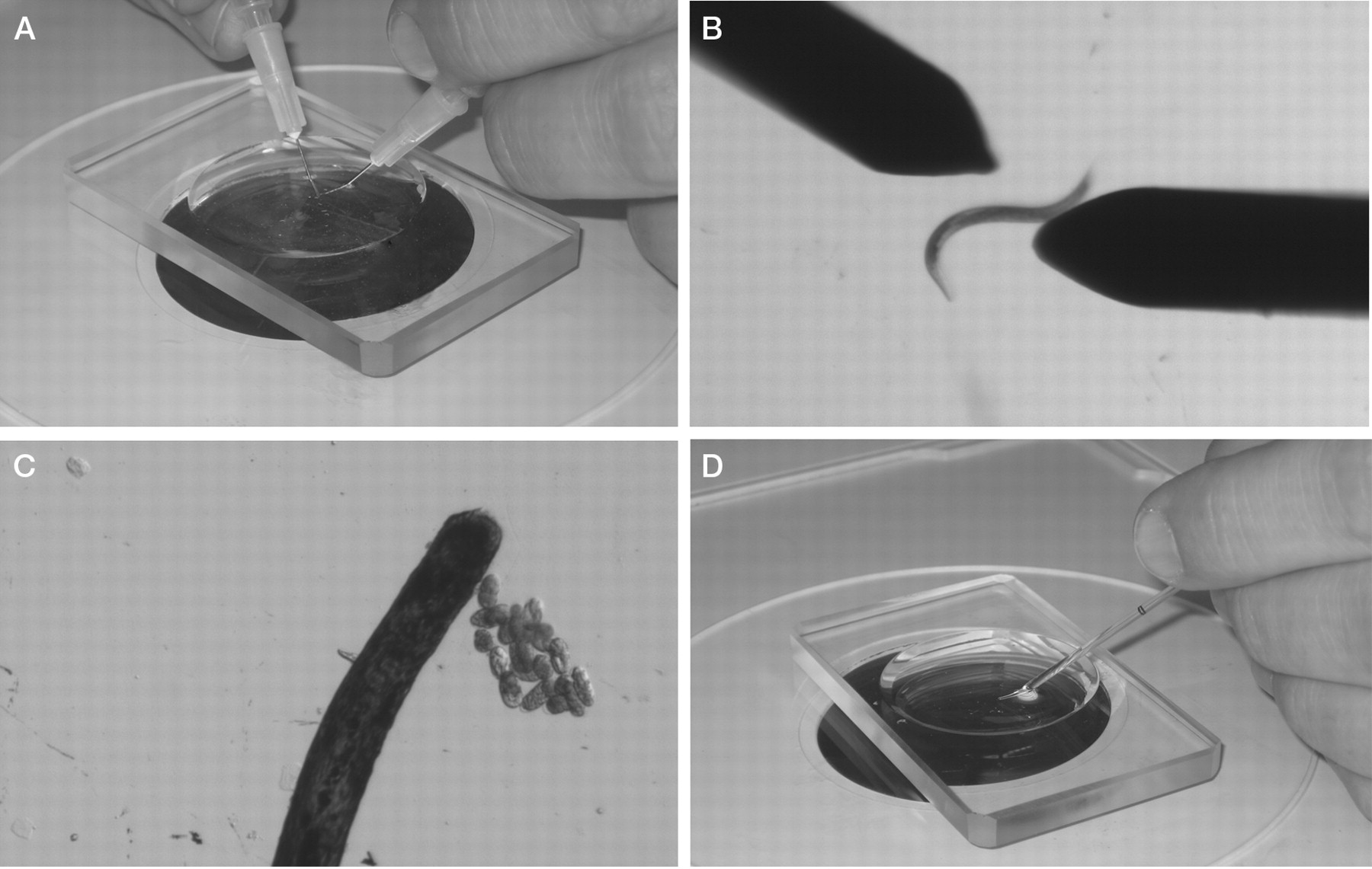
Figure 1.
Isolation of C. elegans embryos and preparation for mounting on a slide. (A,B) Gravid hermaphrodites are cut in half with 27 × 1/2-in. needles. (C) At a higher magnification, embryos are sorted and are grouped using an eyelash. (D) Embryos and M9 buffer are transferred using a mouth pipette.


