
Cover image
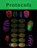
Cover Illustration: Examples of embryos of Caenorhabditis elegans imaged live by confocal and two-photon microscopy. Center 12 images show stereo pairs of two virtually superimposed embryos (one red, one green) undergoing cleavage divisions. Top four images show three distinct developmentally regulated gene expression patterns, both separately and superimposed in false color. Lower left side image shows muscle- and arcade-cell groups expressing lev-8::gfp and aff-1::gfp, each imaged with a 488-nm laser in distinct embryos. Upper left, upper right, lower right, and bottom six images show live embryos labeled with the vital membrane dye FM 4-64. Upper left and lower right show early cleaving embryos in a maximum intensity projection and a single optical section, respectively. (All embryos measure ~50 × 30 µm.) (Images courtesy of William A. Mohler.) For more information on these methods, see Imaging Embryonic Development in Caenorhabditis elegans (doi:10.1101/pdb.top71).

