
Cover image
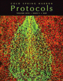
Cover Illustration: Confocal micrograph of the closing neural tube of an intact Xenopus embryo. Image was taken after staining with phalloidin (green) to label actin filaments and propidium iodide (red) to provide contrast. (Image courtesy of Chanjae Lee and John Wallingford.) For more information on these methods, see Preparation of Fixed Xenopus Embryos for Confocal Imaging (doi:10.1101/pdb.prot5426).

