
Cover image
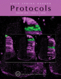
Cover Illustration: Cell tracing by multi-position photoactivation of PSCFP2-labeled cells within the day 4 chick neural tube. Tissue slices (200 μm thick) were cut from the day 4 chick embryo trunk, laid onto a culture insert, and placed in a MatTek dish on a microscope stage surrounded by a heated chamber. Using a Zeiss LSM 510 META confocal microscope equipped with a 10X (0.45 NA) Plan Apochromat and LSM software (Multi-Time), a small region of interest (50 μm in height) was marked and excited with 405-nm laser light that made the photoactivated cells (green) distinct from non-photoactivated cells (purple). Photoactivation was performed sequentially in regions of interest in distinct, neighboring tissue slices (inset) to build a 3D picture of cell movements on one-side of the trunk neural tube. (Image courtesy of Joe Steen, Jennifer Kasemeier-Kulesa, Danny Stark, and Paul Kulesa.) For more information on this method, see Multi-Position Photoactivation and Multi-Time Acquisition for Large-Scale Cell Tracing in Avian Embryos (doi:10.1101/pdb.prot5447).

