
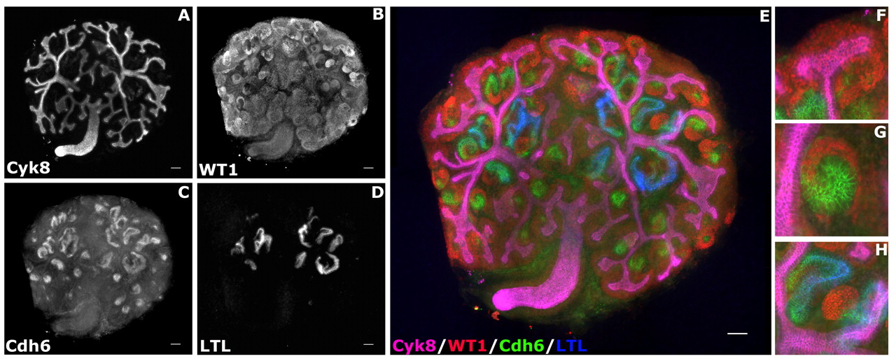
Representative four-color immunohistochemistry of cultured kidneys. (A-D) Black and white images of the four markers used in this study. (A) Cytokeratin 8, ureteric bud/collecting duct. (B) WT1, metanephric mesenchyme, podocytes. (C) Cadherin 6, immature and mature proximal tubules. (D) LTL, mature proximal tubules. (E) Four-color merged image of A-D. (F-H) High-magnification images of various stages of nephron formation. WT1-positive nephron stem cells surround the branched ureteric bud tips (F). These stem cells undergo mesenchyme-to-epithelium transition (MET) to form early nephrons with distinct segments (G). Continued growth and differentiation result in segmented tubular epithelium patterned along a proximo-distal axis (H). Scale bar = 100 microns.


