
Cover image
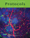
Cover Illustration: Maximum intensity projection of a confocal stack of a mouse mammary carcinoma. Different components of the tumor tissue are visualized in a live MMTV-PyMT;ACTB-ECFP;FSP1EGFP.+/+ mouse using spinning disk confocal microscopy. The cancer cells express ECFP (blue), stromal cells (mostly fibroblasts) express EGFP (green), and blood vessels have been labeled by intravenous injection with rhodamine-dextran (red). For more information on this method, see Dynamic, Long-Term In Vivo Imaging of Tumor-Stroma Interactions in Mouse Models of Breast Cancer Using Spinning-Disk Confocal Microscopy (doi:10.1101/pdb.top97).

