
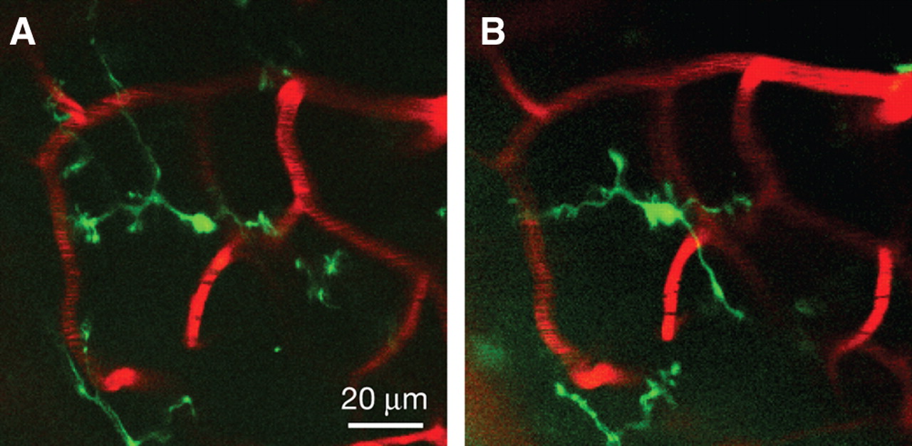
Transcranial TPLSM imaging of enhanced GFP (eGFP)-labeled microglia vasculature within the mouse cerebellum. Two-dimensional projections of a 3D z-stack from the cerebellum (40×, digital zoom = 1) of a mouse harboring a single copy of the CX3CR1-eGFP allele driving eGFP expression in a subset of myeloid cells, including central nervous system resident parenchymal microglia imaged 24 h apart (A,B). The cerebellar vasculature has been labeled in red by intravenous injection of a rhodamine-dextran conjugate solution. The stacks are 6 μm in depth (2-μm step size) and are representative of a section of cortex spanning 40–46 μm below the dural surface. eGFP-labeled microglia retain their characteristic highly branched morphology, indicative of a resting state.


