
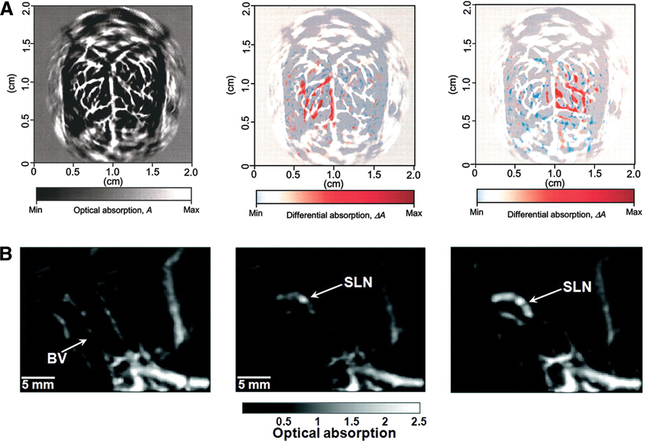
Nontargeted photoacoustic imaging. (A) Functional PAT imaging of cerebral hemodynamic changes in response to whisker stimulation. (Left) A PAT image of the vascular pattern in the superficial layer of the rat cortex acquired with the skin and the skull intact. (Middle) Noninvasive functional PAT images corresponding to left-side whisker stimulation. (Right) Noninvasive functional PAT images corresponding to right-side whisker stimulation. (B) Photoacoustic images acquired before (left), 5 min after (middle), and 140 min after (right) gold nanocage injection for sentinel lymph node (SLN) mapping in live animals. (BV) Blood vessel. (Adapted from Wang et al. 2003b with permission from Macmillan © 2003 and adapted from Song et al. 2009, with permission from American Chemical Society © 2009.)


