
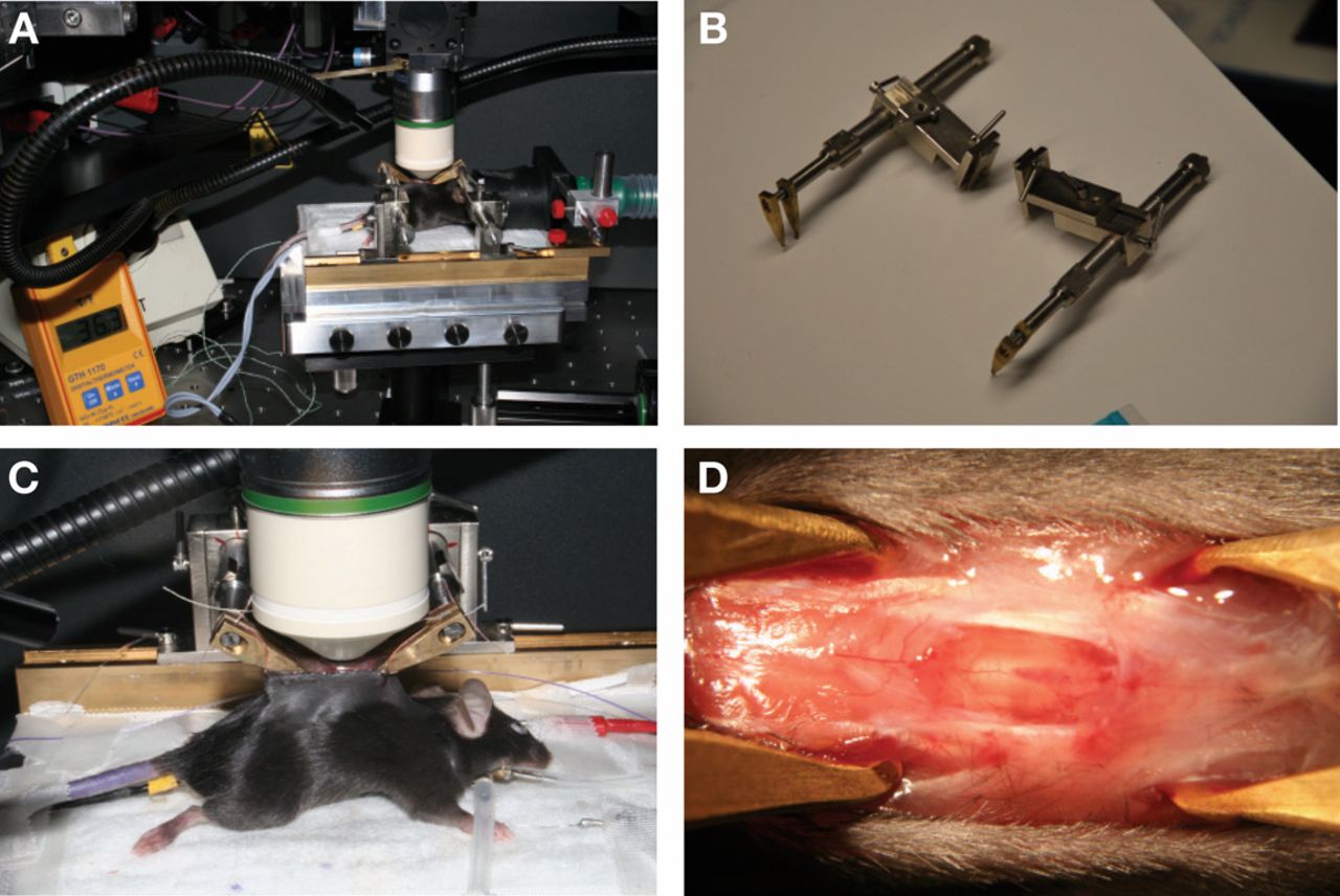
Figure 1.
Preparation of mice for spinal cord imaging. (A) Mouse receives a volatile anesthetic, a rectal probe (see also C) monitors temperature, and imaging is performed with a high numerical aperture, 20× objective. (B) Clamps to fix the vertebral spines. (C) Mouse is artificially ventilated through a tracheal tube for single imaging. Clamps fix the vertebral column. Skin flaps form a reservoir for artificial cerebrospinal fluid (ACSF) in which the objective is immersed. (D) Exposed spinal cord after a laminectomy.


