
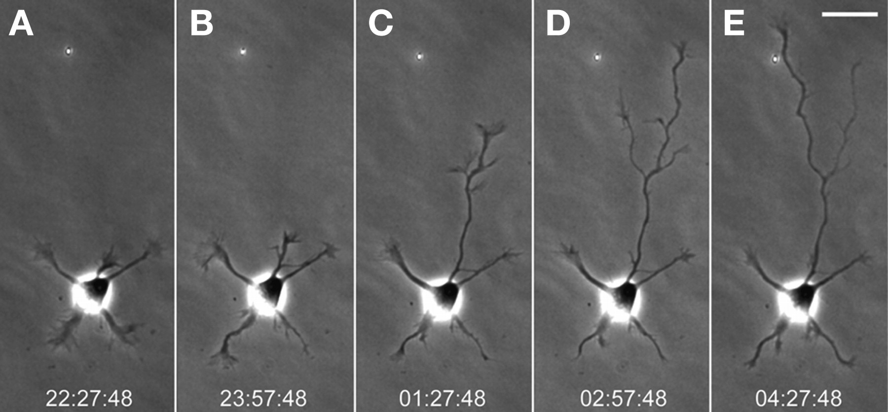
Long-term imaging of a neuron during axonal specification. These frames were taken from a time-lapse recording in which images were acquired at 10-min intervals over a period of 13 h (see Movie 1). A fully automated wide-field microscope was used together with software-based autofocusing. During stage 2 (A,B), minor neurites extended and retracted but showed little net growth. In the next interval (C), one neurite became longer than the rest; at this point in development, the identity of the axon is certain. In the final two intervals (D,E), the axon extended still farther and gave off a collateral branch. The branch became dominant and extended quickly while the initial process stalled. Time stamps show h:min:sec. Scale bar, 25 µm.


