Cover image
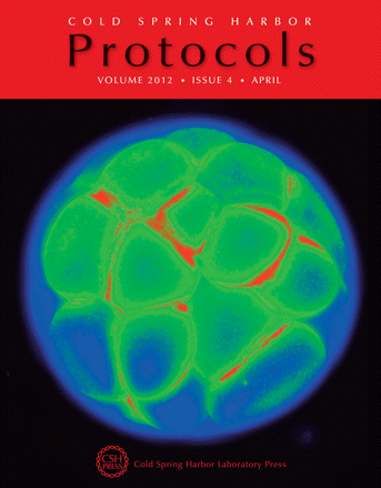
A stage 6 (32-cell) Xenopus laevis embryo imaged using the voltage-sensitive dye DiBAC4(3). For an introduction to the use of fluorescent bioelectricity reporters, see the article by Adams and Levin in this issue (Cold Spring Harb Protoc 2012; doi: 10.1101/pdb.top067710); for a method using DiBAC4(3) along with CC2-DMPE to image membrane voltage in Xenopus embryos, see the related protocol by Adams and Levin (Cold Spring Harb Protoc 2012; doi: 10.1101/pdb.prot067702). Image taken with a BX-61 Olympus Scope, 4× lens, epifluorescent optics, using the FITC filter set (470/20, 485, 517/23; courtesy of Dany Adams).