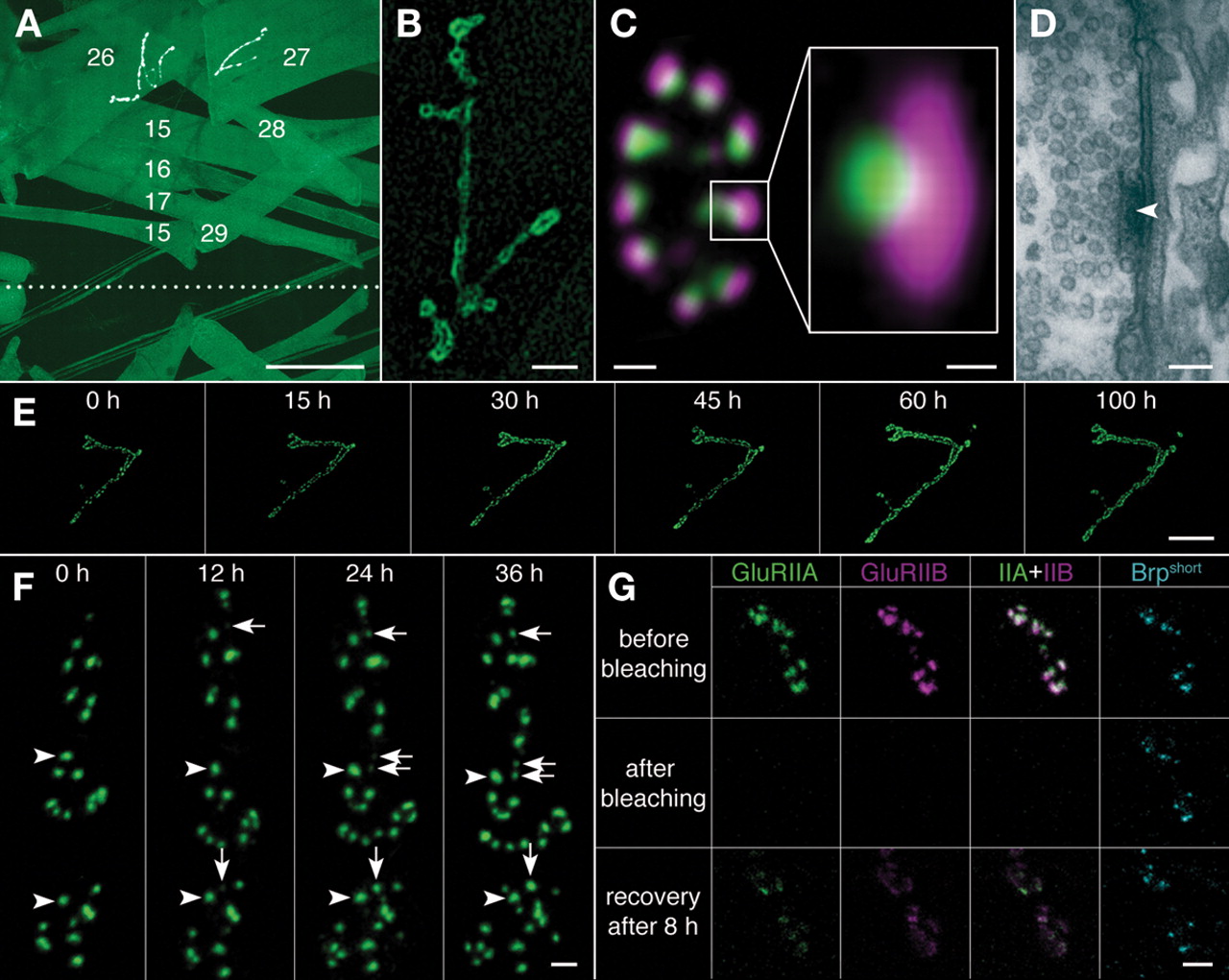
Intravital imaging of neuromuscular junctions (NMJs). (A) Selected body wall muscle fibers in an abdominal segment as seen from the exterior (outside-out view). The dotted line indicates the ventral midline. The image was modified to highlight NMJs at muscles 26 (VA1) and 27 (VA2). Construct used for the visualization of muscle fibers and NMJs: uas-dlgS97-gfp, expressed using c57-gal4. Scale bar, 100 µm. (B) Morphological structure of a Drosophila larval NMJ at muscle 27 (VA2), outlined by the expression of uas-dlgS97-gfp, using c57-gal4 as a driver. Scale bar, 10 µm. (C) Immunohistochemical stainings of a bouton of a larval NMJ and an individual synapse in lateral view (right box). Green, monoclonal antibody BruchpilotNc82 (Kittel et al. 2006); magenta, antibody against the glutamate receptor subunit DGluRIID (Qin et al. 2005). Scale bar (bouton), 1 µm; scale bar (synapse), 100 nm. (D) Ultrastructure of an active zone. The arrowhead points at the T-bar, synaptic vesicles cluster next to it. Scale bar, 100 nm. (E,F) In vivo imaging of synapse formation at an identified NMJ at muscle 27, tracked using the GFP-labeled glutamate receptor subunit DGluRIIA. (Adapted, with permission, from Rasse et al. 2005.) (E) The development of an identified NMJ at muscle 27 tracked >100 h at 16°C. Scale bar, 10 µm. (F) Higher magnification of the image shown in E: New synapses form de novo (arrows). Mature synapses (arrowheads) remain stable. Scale bar, 2 µm. (G) In vivo fluorescent bleach experiment (FRAP) of DGluRIIA-mRFP (green), DGluRIIB-GFP (magenta) and Brpshort-Cerulean (cyan). (Top row) Images taken directly before bleaching. (Middle row) Images taken directly after bleaching of DGluRIIA and DGluRIIB. (Bottom row) Images taken 8 h after bleaching. Recovery of both DGluRIIA and DGluRIIB can be observed. Scale bar, 2 µm.