
Cover image
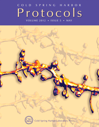
Nanoscale imaging of living synapses. This image of dendritic spines on a hippocampal neuron inside a brain slice was acquired on a home-built stimulated emission depletion (STED) microscope using yellow fluorescent protein (YFP) as a volume label and volume-rendered in pseudo-color using the Imaris software. See Willig and Nägerl (Cold Spring Harb Protoc 2012; doi: 10.1101/pdb.prot069260). Image courtesy of U. Valentin Nägerl.

