
Cover image
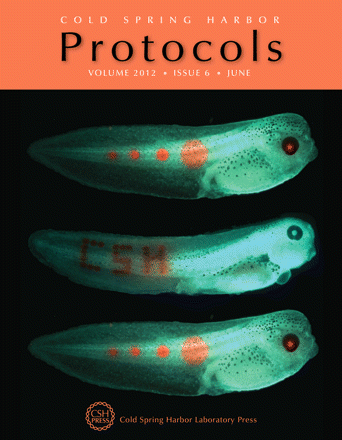
The protocol by Chernet et al. (2012; doi: 10.1101/pdb.prot068502) in this month's issue allows the user to convert green-labeled cells to a red color with great spatial specificity. Here, this technique was used to photoconvert cells in a frog embryo into a pattern that reads “CSH.” This technique is useful for tracking the fate and positioning of key cell groups during embryogenesis, regeneration, or cancer progression. (Tadpole images courtesy of Brook Chernet.)

