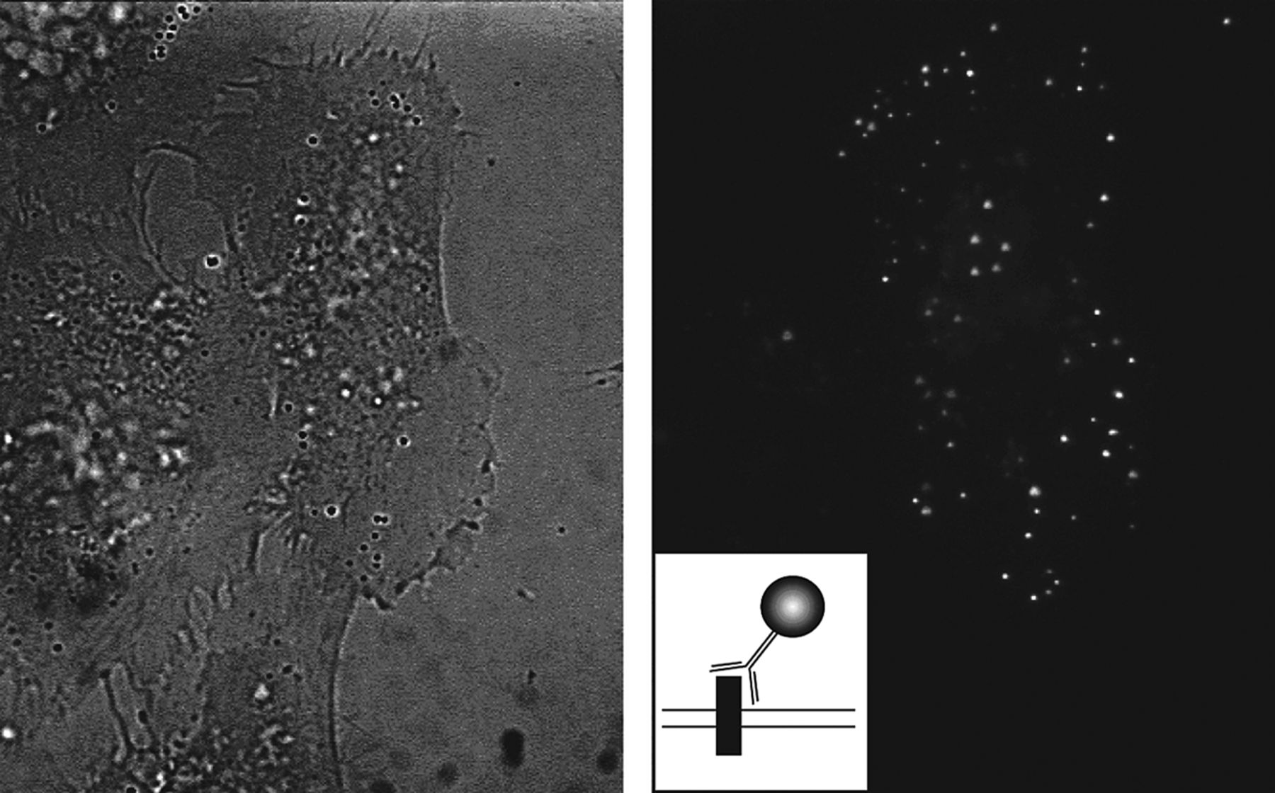
Figure 1.
Example of surface QD labeling on transfected cells. (Left) Bright-field image of cultured HeLa cells. (Right) Fluorescence image. Only one of the cells is transfected with a membrane receptor possessing an extracellular myc tag. Individual spots correspond to receptors labeled with anti-myc antibodies and QDs (see inset).