
Cover image
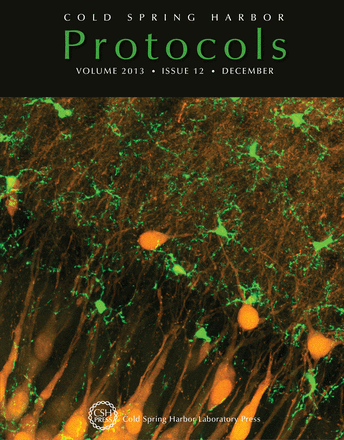
Confocal image stack showing GFP-expressing microglia (green) and YFP-expressing neurons (yellow) in area CA3 of a hippocampal tissue slice from postnatal day 12 mouse (CX3CR1GFP/+:Thy1-YFP). In this issue, Michael Dailey and colleagues describe how to image fluorescently labeled microglia in neonatal and adult rodent brain tissue slices (doi: 10.1101/pdb.prot079483). Image courtesy of Michael E. Dailey.

