
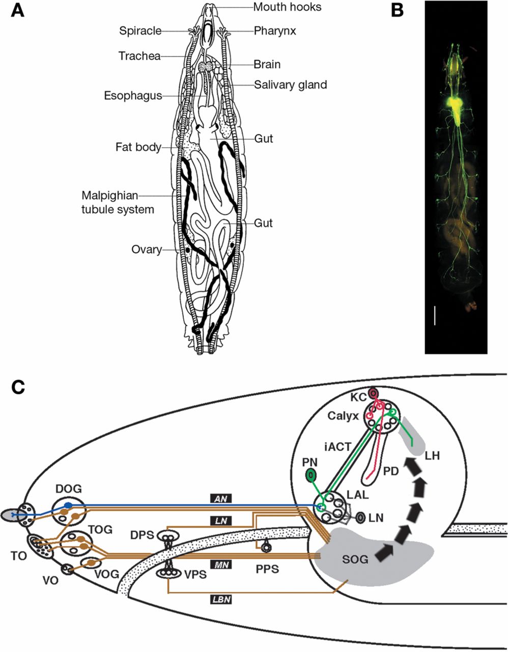
(A) Major body parts of a Drosophila larva. (B) Larval nervous system. Fluorescent image of the larval nervous system, clearly showing the brain with the ventral nerve cord and the major nerve cords. Scale bar, 100 μm. (C) Circuitry. Overview of the chemosensory pathways of the Drosophila larva. Olfactory pathways (blue) project into the brain proper, whereas gustatory input (brown) is collected in various regions of the subesophageal ganglion. The bold arrows indicate the proposed octopaminergic pathway to short circuit a taste-driven “value” signal carried by neurons from the subesophageal ganglion toward the brain. (Reprinted, with permission of Landes Bioscience, from Stocker 2008.) AN, antennal nerve; DO/DOG, dorsal organ/ganglion; DPS, dorsal pharyngeal sense organ; iACT, inner antennocerebral tract; KC, Kenyon cells; LAL, larval antennal lobe; LBN, labial nerve; LH, lateral horn; LN, local (inter)neurons; LN, labral nerve; MN, maxillary nerve; PD, pedunculus; PN, projection neuron; PPS, posterior pharyngeal sense organ; SOG, subesophageal ganglion; TO/TOG, terminal organ/ganglion; VO/VOG, ventral organ/ganglion; VPS, ventral pharyngeal sense organ. (A) Courtesy of The Carnegie Institution; (B) reprinted, with permission, from Sun et al. 1999 (© National Academy of Sciences, USA); (C) modified, with permission from Landes Bioscience, from Stocker 2008.


