
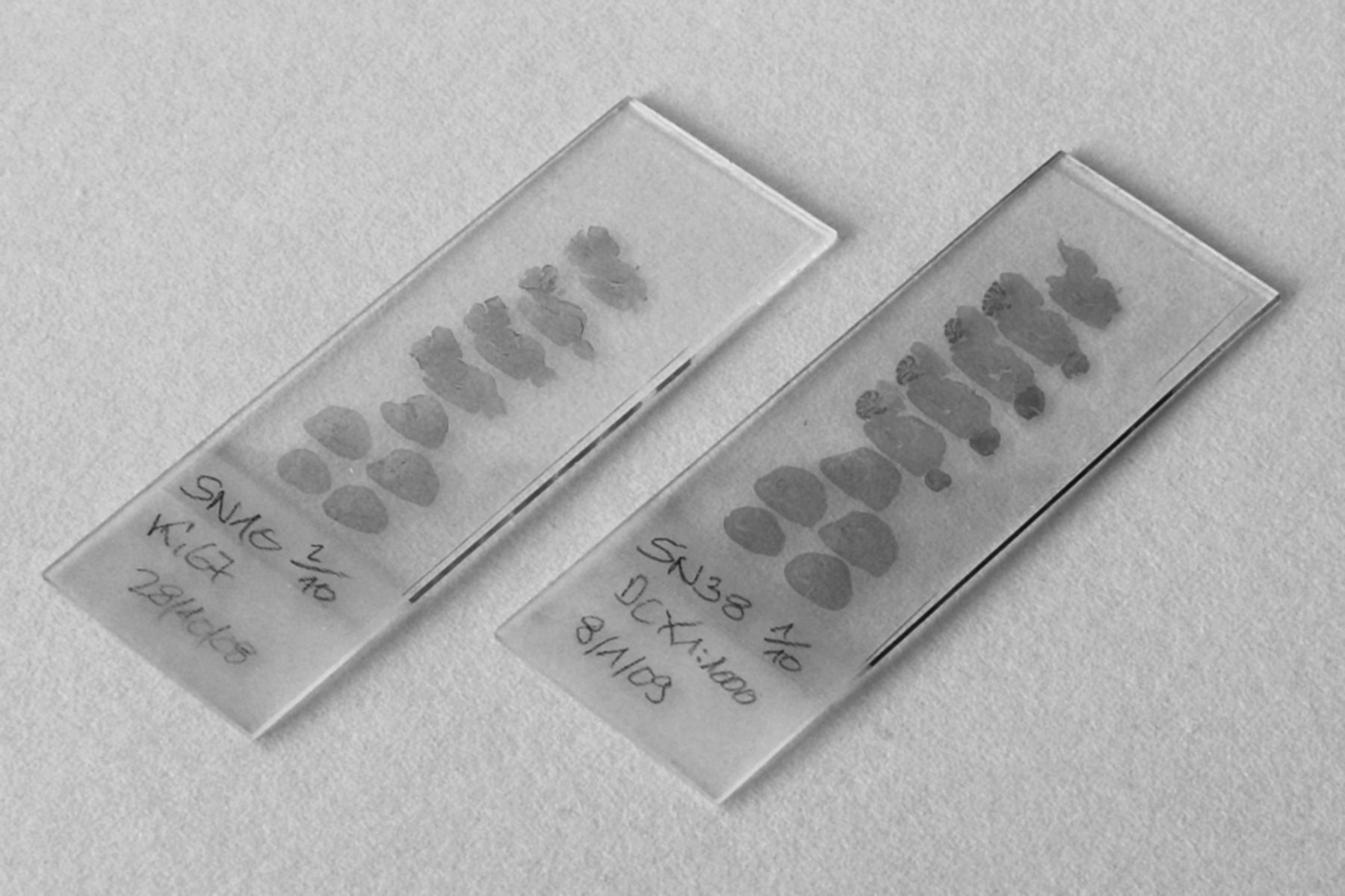
Figure 1.
Two immunocytochemically stained series (left, proliferation marker Ki67; right, microtubule associated protein doublecortin) of mouse brain sections containing hippocampus. Sagittal sections were cut frozen at 40 μm, subjected to antigen retrieval, and stained free-floating. No sections or parts of sections were lost from either series. Note that the top-right section of both series looks incomplete. They are the last sections cut from the hemispheres, in which parts of other structures already had disappeared. Sections of this type also need to be collected if they contain part of the region of interest. (Courtesy of Lutz Slomianka.)


