
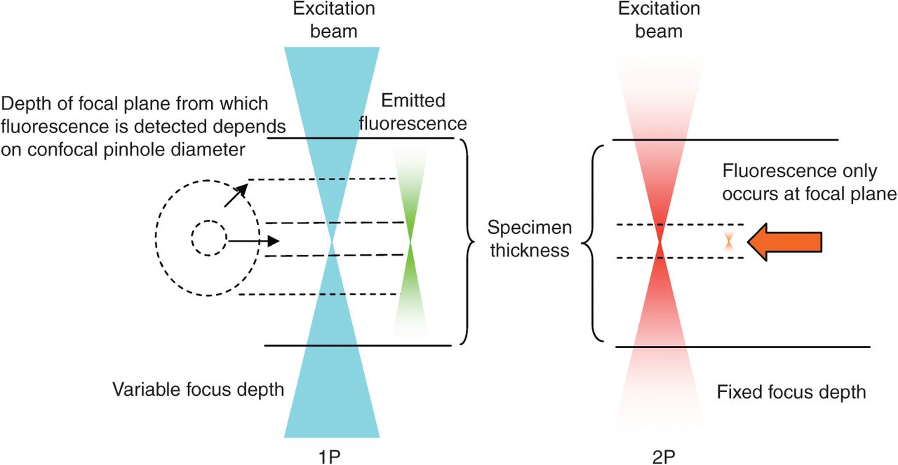
Figure 5.
Determination of depth-of-field and bleaching in one- or two-photon microscopy. In one-photon microscopy (1P, blue), a cone of excitation light on either side of the focal point illuminates and excites the fluorescence indicator throughout the specimen. The size of the pinhole determines the depth of fluorescence detection. In two-photon microscopy (2P, red), the photon density is only sufficiently high at the focal plane to induce indicator fluorescence. This beam has a fixed depth of excitation.


