
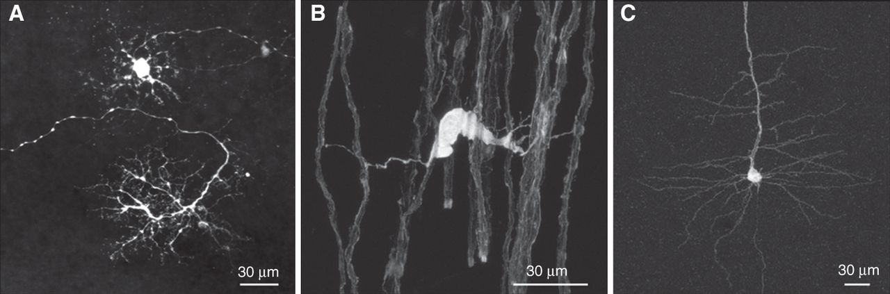
Figure 3.
Examples of infection of diverse cells types by SAD ΔG-eGFP (G). (A) Horizontal cells in an explanted mouse retina. (Courtesy of Botond Roska, Friedrich Miescher Institute, Basel.) (B) Oligodendrocyte after injection of SAD ΔG-eGFP (G) into the spinal cord. (C) Layer V neuron in neocortex. (B,C, Courtesy of Martin Kerschensteiner, Ludwig-Maximilians-University, Munich and Thomas Misgeld, Technical University, Munich.)


