
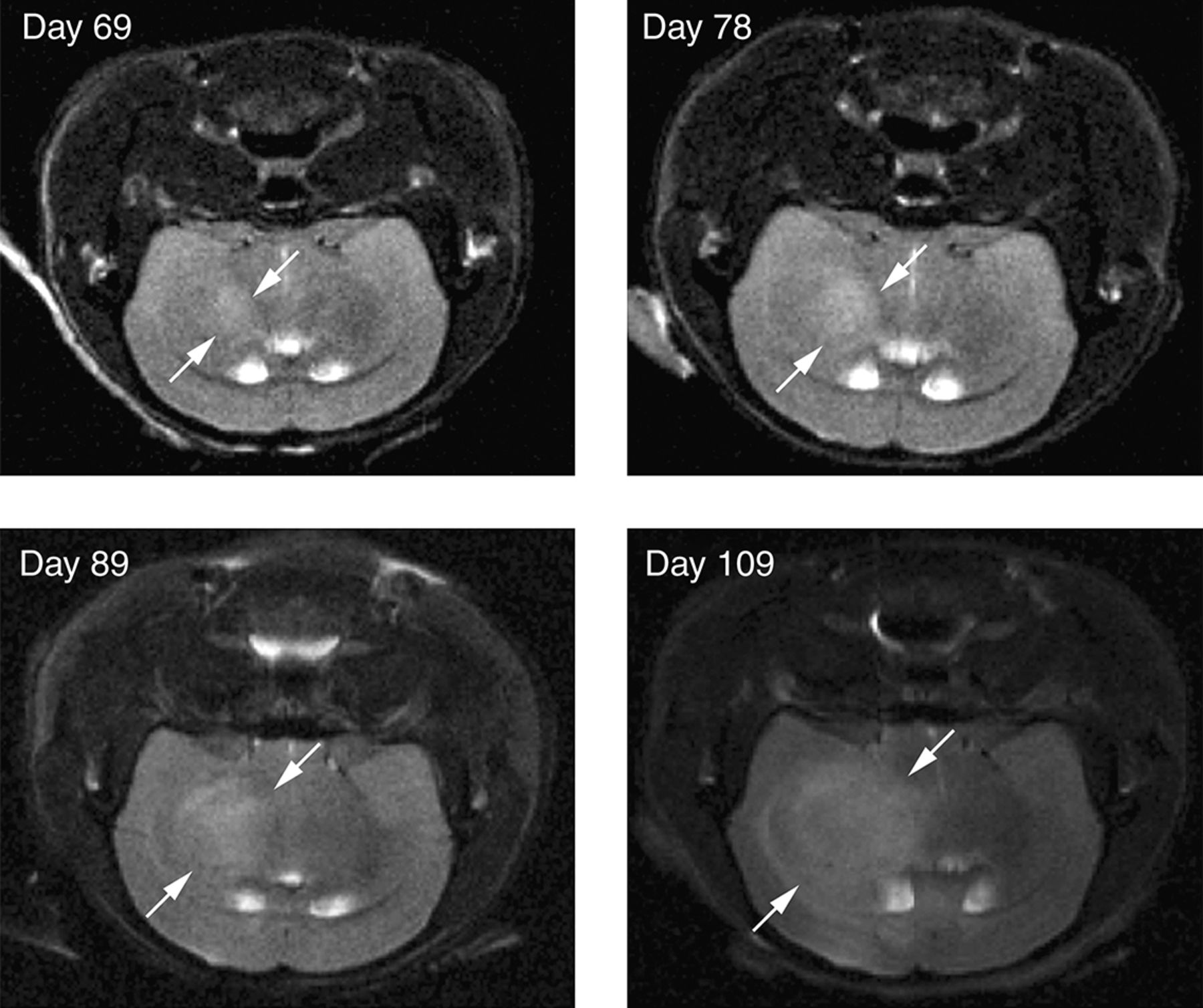
Figure 2.
MRI of orthotopic brain tumors. Primary human glioblastoma multiforme cells were stereotactically implanted into the brains of mice. MRI imaging shows progressive growth of primary orthografts at the indicated time after implantation. The tumor (boundary indicated by arrows) is distinguishable from surrounding brain tissue on the basis of T2 hyperintensity (transverse relaxation).


