
Cover image
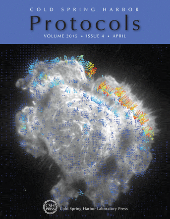
Velocity vector map of α-actinin-mCherry in a CHO.B2 cell calculated using spatiotemporal image correlation spectroscopy (STICS). The vector map is superimposed on the first image of the time window analyzed (frames 45–54 of duration 50 sec in total). Pixel size is 0.105 µm and each STICS calculated region of interest is 16×16 pixels (1.7 µm×1.7 µm). In this issue, Paul Wiseman introduces various versions of image correlation spectroscopy, including STICS (see doi: 10.1101/pdb.top086124). STICS analysis by Tim Toplak and Prof. Paul W. Wiseman of McGill University. Video microscopy collection by Dr. Miguel Vicente Manzanares and Prof. A. Rick Horwitz of the University of Virginia.

