
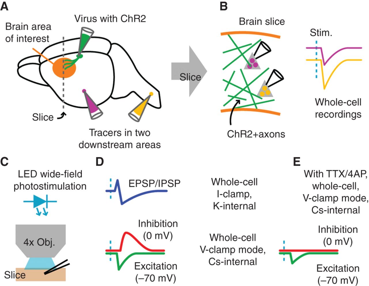
General paradigm for optogenetic circuit electroanatomy. (A) In vivo injections are made to deliver either rabies or adeno-associated virus carrying ChR2 to one brain region of interest that is either upstream or downstream from the main region of interest (orange area indicates where brain slices will be made), and to deliver retrograde tracers of two different colors in areas that are downstream from this area. (B) In brain slices of the main brain area of interest, ChR2-expressing presynaptic axons (green) are photostimulated while recordings are made from the two types of projection neurons identified by fluorescent labeling. (C) LED illumination through a low-power objective (Obj.) is used for wide-field photostimulation. (D) Electrophysiological manipulations allow different types of responses to be sampled, including mixed excitatory and inhibitory postsynaptic potentials (EPSP/IPSP) in current-clamp (I-clamp) mode, or isolated inhibitory and excitatory currents in voltage-clamp (V-clamp) mode. “K-internal” and “Cs-internal” refer to the cation used in the internal (pipette) solution; see Table 1. (E) Pharmacological manipulations allow isolation of purely monosynaptic inputs.


