Cover image
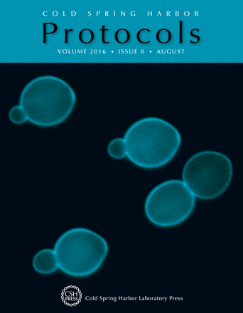
Fluorescent image of wild-type (BY4743) Saccharomyces cerevisiae cells stained with aniline blue to detect the cell wall component 1,3-β-glucan. A protocol for staining and visualizing S. cerevisiae cell wall components is included in this issue (doi: 10.1101/pdb.prot085241). Image courtesy of Hiroki Okada and Yoshikazu Ohya.