
Cover image
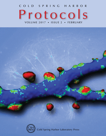
Three-dimensional reconstruction of a live CA1 pyramidal cell dendrite in rat hippocampal slice culture, demonstrating enrichment of αCaMKII in dendritic spines. The colors represent the ratio of αCaMKII-GFP fluorescence to volume-filling RFP fluorescence (see Zhang et al. 2008; Proc Natl Acad Sci 105: 12039–12044). A protocol for the preparation of organotypic slice cultures is included in this issue (doi: 10.1101/pdb.prot094888). The image was generated by Yanping Zhang in the laboratory of Thomas Oertner at the Friedrich Miescher Institute in Basel.

