
Cover image
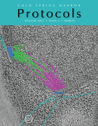
A complete 3D model of a mitotic spindle from budding yeast (spindle microtubules, green and pink lines; spindle pole bodies, blue discs; and cytoplasmic microtubules, teal lines) projected on an electron tomographic slice. In this issue, Eileen O'Toole, Thomas Giddings, and Mark Winey discuss electron tomography methods for budding yeast, emphasizing the advantages of preparing the cells by high-pressure freezing and freeze substitution (doi: 10.1101/pdb.top077685). Image courtesy of Eileen T. O'Toole (University of Colorado Boulder).

