
Cover image
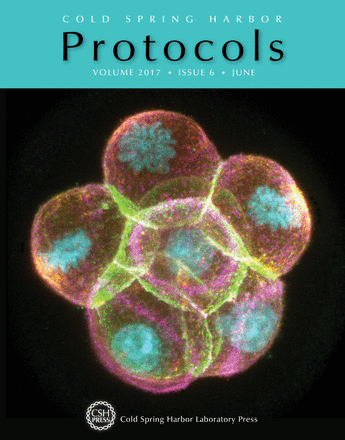
Eight-cell preimplantation embryo undergoing cleavage (E-cadherin, green; F-actin, pink; phospho-myosin II, red; and DNA, blue). In this issue, Yojiro Yamanaka describes how to construct a convenient chamber for observing mouse embryos under the microscope (doi: 10.1101/pdb.prot094045). Image courtesy of Deepak Saini in the laboratory of Yojiro Yamanaka, McGill University.

