
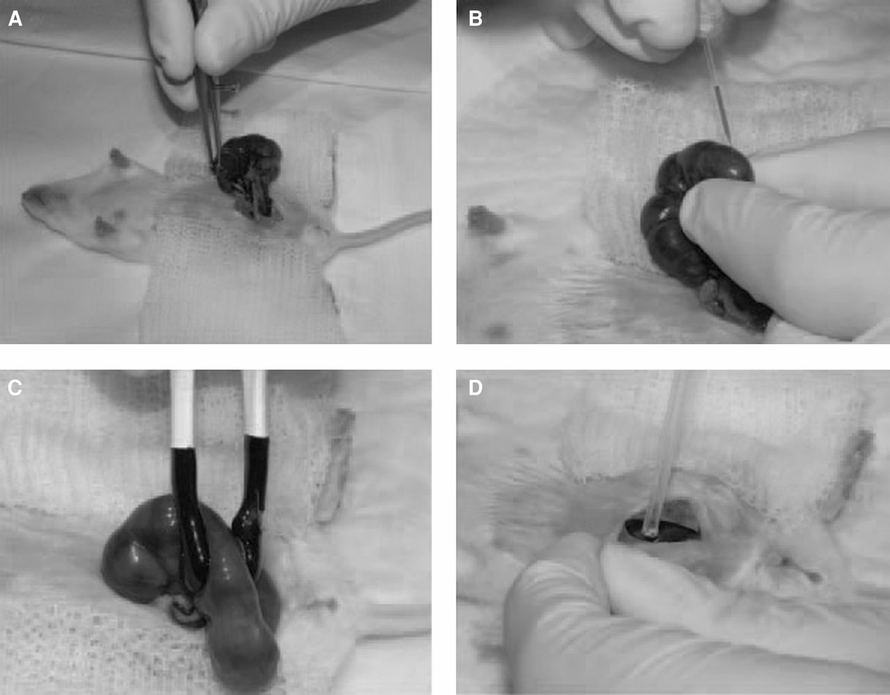
Figure 1.
In utero electroporation of gene constructs. (A) Anesthetized pregnant female mouse with abdominal incision. The uterine horn with fetuses is gently pulled outside of the body. (B) A fetus within the uterus is cradled by hand, and a DNA solution in a pulled glass capillary is injected into the brain ventricle. (C) Electroporation paddles are placed on the uterus flanking the injected fetus, and electrical pulses are given. (D) The uterus is placed back in the abdominal cavity.


