
Cover image
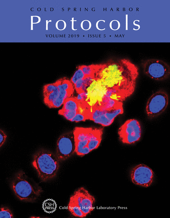
Confocal fluorescence micrograph of peritoneal leukocytes harvested from Xenopus laevis tadpoles infected with green fluorescent Mycobacterium marinum for 4 h in vitro. Cell membranes were then labeled with PKH (red), and nuclei were labeled with Hoescht (blue). In this issue, Kun Hyoe Rhoo and Jacques Robert provide a protocol for adoptive cell transfer in Xenopus to study immune processes in real time (doi: 10.1101/pdb.prot097592). Image courtesy of Jacques Robert.

