
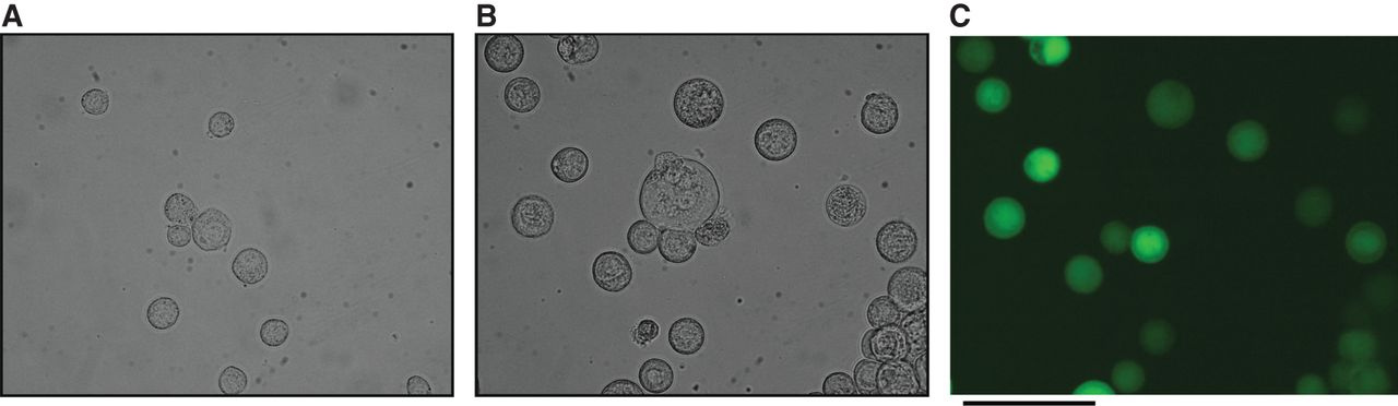
Figure 1.
Changes in the appearance of baculovirus–infected insect cells. Tn-5 cells uninfected (A) and infected (B,C) with baculovirus generated using a pVL1393-based transfer vector and linearized ProGreen genomic baculovirus vector DNA. The infected cells express GFP encoded in the ProGreen DNA. Panel C is a fluorescence image of the same cells as shown in B with phase-contrast illumination. Note that the infected cells are on average slightly larger than the uninfected cells. Scale bar, 100 µm. (Photographs courtesy of Drs. Jost Vielmetter and Inderjit Nangiana, Caltech Protein Expression Facility.)


