
Nonlethal Blood Sampling from the Killifish Nothobranchius furzeri
- Luca Dolfi1,3,
- Roberto Ripa1,3,
- Danel Medelbekova1,
- Eugen Ballhysa1,2,
- Orsolya Symmons1 and
- Adam Antebi1,2,4
- 1Max Planck Institute for Biology of Ageing, 50931 Cologne, Germany
- 2Cologne Excellence Cluster on Cellular Stress Responses in Aging-Associated Diseases (CECAD), University of Cologne, 50923 Cologne, Germany
- ↵4Correspondence: aantebi{at}age.mpg.de
-
↵3 Equal contribution
Abstract
Blood withdrawal is a common procedure performed on laboratory animals to monitor key processes and indicators of fish health and physiology, such as hematopoiesis, hemostasis, and lipid and glucose metabolism. Moreover, the ability to extract blood with minimal invasiveness and without sacrificing animals enables repeated sampling, allowing both longitudinal studies of individual animals, as well as reducing the number of experimental animals needed in a study. The African turquoise killifish is an emerging animal model that is progressively being adopted worldwide for aging studies because of its naturally short life span. However, because of the small body size of this species, nonlethal blood collection is a challenging procedure. Here we present a detailed protocol enabling repeated blood sampling from the same individual fish. This method, if correctly executed, is minimally invasive and does not cause any lasting damage. The protocol has been tested on animals spanning from 6 to 24 wk of age and the amount of blood that could be extracted varied from 0.5 to 8 µL, greatly depending on specimen age, sex, and size. This volume is sufficient to perform analyses such as blood glucose measurement, blood cell counts, or histological stains on blood smears.
MATERIALS
Reagents
Aquarium H2O
-
In our laboratory we use chlorine-free fresh H2O with pH 7.0–7.2, salinity of 650–710 µS/cm, and temperature 27.0°C–27.5°C. However, given that H2O conditions for killifish can vary widely in their natural habitat, other parameters can also be acceptable (Polačik et al. 2016).
Dulbecco's Phosphate Buffered Saline (DPBS; Gibco 14190-094)
Heparin solution (Sigma-Aldrich 9041-08-1; 3% w/v in DPBS), freshly prepared and sterilized
Nothobranchius furzeri >6 wk raised according to Dodzian et al. (2018)
Tricaine methanesulfonate (ethyl 3-aminobenzoate methanesulfonate; Sigma-Aldrich E10521) (0.5 mg/mL in aquarium H2O)
Equipment
Aquarium fish net
Aspirator tube assembly 15″ latex with connector (Alpha Laboratories 2-000-001)
Block of modeling clay
Forceps
Glass capillaries (Warner Instruments 64-0766)
Glucometer (Accu-Chek) and test strips (optional; see Step 17)
Microcentrifuge tubes (1.5-mL)
Needle puller (Tritech PP-830 or similar)
Paper towels
Petri dish (10-cm)
Rubber pipette bulb
Small plastic/glass box (∼0.5-L)
Stereomicroscope with top illumination
Vessel, glass slides, or Parafilm for collecting the blood sample
METHOD
-
Assistance from another person is not necessary but may be helpful to minimize handling times and thereby reduce stress to animals and avoid changes to blood parameters. We particularly recommend using an additional person if the number of specimen is substantial (>10) and/or the amount of time available for the procedure is limited (<2 h). We recommend dividing the work as follows: One person prepares the needle (inserting the needle in the aspirator, breaking the tip of the needle, and heparinization) and executes the blood draw, the other person prepares the fish for the procedure, and recovers the blood into a collection tube or onto a slide or Parafilm.
-
See Figure 1A for a picture of the equipment needed for blood extraction.
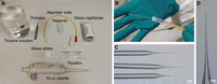
(A) Equipment for blood extraction. (B) Needle assembly on the aspirator tube. (C,D) Example of pulled needles (C) and tip opening (D) required for the procedure.
-
1. Using a needle puller (Tritech PP-830 model, set to one step at 58.5°C), pull the required number of needles (typically 1.5× number of fish + 5) from glass capillaries. Store the needles temporarily in a large Petri dish on a clay block. Make needles similar in shape to the ones pictured in Figure 1C.
-
2. Place 1 mL of 3% w/v fresh sterile heparin solution in DPBS in a 1.5-mL microcentrifuge tube.
-
3. Under a dissecting microscope, carefully break off the needle to approximately one-third of the tip, using forceps (Fig. 1D).
-
4. Attach the needle to one end of the rubber aspirator tube and generate an airtight seal by pushing 0.5–1 cm of the glass capillary inside (Fig. 1B).
-
5. Heparinize the needle to avoid coagulation of blood, by applying negative pressure to the aspirator tube using a rubber pipette bulb. Place the needle in the microcentrifuge tube with heparin and pipette the heparin up and down within the glass capillary several times to coat the inside. At the end, gently release all the heparin, leaving the needle empty.
-
Note that the flow of heparin should be smooth. If there is excessive resistance, open the needle tip further.
-
-
6. Fill a plastic box with tricaine methanesulfonate solution (0.5 mg/mL in 4°C refrigerated aquarium water). Using a net, immerse the fish to anesthetize.
-
Anesthesia with tricaine methanesulfonate solution can potentially modify blood parameters that change rapidly with the environment. This should be considered when planning experiments. (Carter et al. 2011; Larter and Rees 2017) provide some additional information on how tricaine methanesulfonate anesthesia can impact physiological parameters in other species.
-
-
7. When the fish stops swimming and opercular movement is greatly reduced (approximately one opening every 60 sec), collect the fish with the net and lay it on top of a paper towel under the microscope, with its head pointing to the left (for right-handed experimentalists) or to the right (for left-handed experimentalists), with the dorsal fin pointing away from the researcher.
-
The operculum is a bony plate that covers the fish's gills and serves as a water pump. Observation of the opercular movement allows evaluation of the respiration rate.
-
-
8. Thoroughly dry the region from the pectoral fin to the caudal fin using a paper towel, as shown in Figure 2A.
-
9. Use forceps to remove one scale from the skin at the lateral midline, at the same vertical position as the posterior end of the dorsal fin (Fig. 2B,C).
-
10. Gently press the needle to the skin spot where the scale was removed, forming an angle of ∼60° with the surface (Fig. 2D).
-
11. While maintaining negative pressure with the micropipette, slowly press the needle through the skin, keeping the direction of penetration close to the midline in order to reach the dorsal aorta without touching the bones (to avoid spinal damage).
-
12. To retrieve the blood, use the hand holding the needle to detect two click points of penetration—the first upon breaking the skin and entering the muscle, and the second upon entering the dorsal aorta. Immediately after the second click, blood should start to flow rapidly into the needle without any suction (Fig. 2E).
-
If blood does not flow, gently move the needle around until the blood vessel is found.
-
-
13. Keep the needle immobile and let it fill with blood. Upon reaching the desired volume, release negative pressure in the micropipette to stop the blood flow.
-
The maximum volume of blood sampling is dependent on the body weight of the fish. Although there are no exact recommendations for acceptable blood volumes, we suggest extracting no >10% of blood volume, which is∼3–4 µL of blood from fish with ≤1.0 g body weight and up to 6–8 µL from fish with ≥2.0 g body weight. For further reference on blood volume considerations we also recommend (Lawrence et al. 2020).
-
-
14. Gently extract the needle and transfer the fish back into clean, warm (27°C) aquarium water for recovery.
-
15. Carefully use forceps to completely break off the needle tip.
-
16. Recover the blood by positioning the end of the needle into a suitable vessel (microcentrifuge tube) or onto a suitable surface (e.g., Parafilm, glass slide) and subsequently apply positive pressure to release the contents.
-
The collected blood can be used for a number of downstream applications such as: glucose blood test, blood smear examination, nucleic acids extraction, or immunohistochemistry analysis (Zang et al. 2015).
-
-
17. If measuring blood glucose levels, immediately use 1.5–2 µL of blood for this purpose. Release the blood onto a piece of Parafilm or a coverslip. Touch the blood with the strip and then insert the strip into a commercially available glucometer. Observe the reading on the glucometer (Fig. 3A–C).
-
Use the blood right after the extraction to avoid coagulation. Do not dilute the blood with DPBS or other solutions before measurement.
-
-
18. Safely dispose of the needle and prepare a new one for the next fish.
-
19. Check that the first fish has regained consciousness within a few min. Bleeding is typically minimal and no bleeding should be observed after 5 min following the blood draw. In the following days, house the fish individually to allow recovery and healing. Assess for infection or motility problems daily. The needle wound should heal completely within a week.
-
20. Repeated extractions from the same fish are possible; however, intervals of <1 wk are not recommended to avoid unnecessary stress on the animal. Ideally, the next extraction should be performed after 1 mo or more.
-
Repeated extractions can target exactly the same locus (which will appear cicatrized in most cases and therefore easy to identify) or the opposite side of the fish. After a successful extraction we recommend targeting the same spot at the next time point because the scar will guarantee the correct position of the vessel, and most likely the success of the subsequent blood withdrawal. In case the extraction was problematic or not smooth we recommend changing sides for the next time point.
-
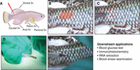
Blood extraction procedure. (A) Schematic representation of the killifish anatomic landmarks. (B) Anatomical area suitable for the needle insertion. (C) Scale removal from the chosen spot. (D) Needle insertion at a ∼60° angle with the surface. (E) Blood flowing into the needle.
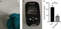
Blood glucose measurement: (A) Collected blood is rapidly recovered on a glass slide or parafilm. (B) Representative glucometer commercially available. (C) Blood glucose levels in fully fed and 48 h fasted fish (n = 6 fish per experimental group, P = 0.0079, Mann–Whitney U-test).
ACKNOWLEDGMENTS
This project has received funding from the European Research Council (ERC) under the European Union's Horizon 2020 research and innovation programme (grant agreement No 834259).
Footnotes
-
From the African Turquoise Killifish collection, edited by Anne Brunet.


