
Cover image
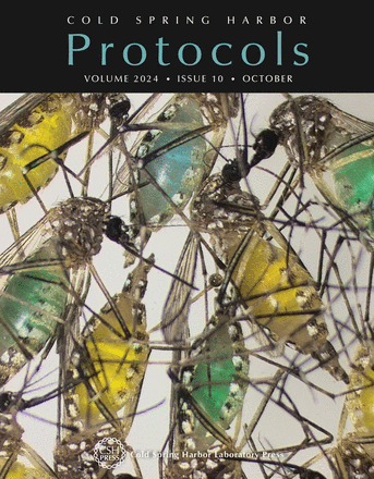
Mosquitoes are ideal model organisms for studying egg maturation in arthropods, as their follicle development is initiated only after the ingestion of a blood meal. The amount of blood consumed is a key factor impacting fecundity and the fate of maturing oocytes, as small blood meals—and the consequent nutrient limitation—may result in a significant fraction of follicles undergoing apoptosis and oosorption. Suitable methods for the evaluation of follicular atresia are needed to understand the mechanisms underlying follicle development in insects. In this issue, Isoe et al. describe an efficient imaging approach for visualizing apoptotic ovarian follicles during Aedes aegypti egg maturation (doi:10.1101/pdb.prot108226). The cover image shows Aedes aegypti female mosquitoes that were fed an artificial protein diet that was spiked with a food dye, which can be seen in the midguts of engorged mosquitoes and allows monitoring of feeding activity under various experimental conditions. Image provided by Jun Isoe.

