
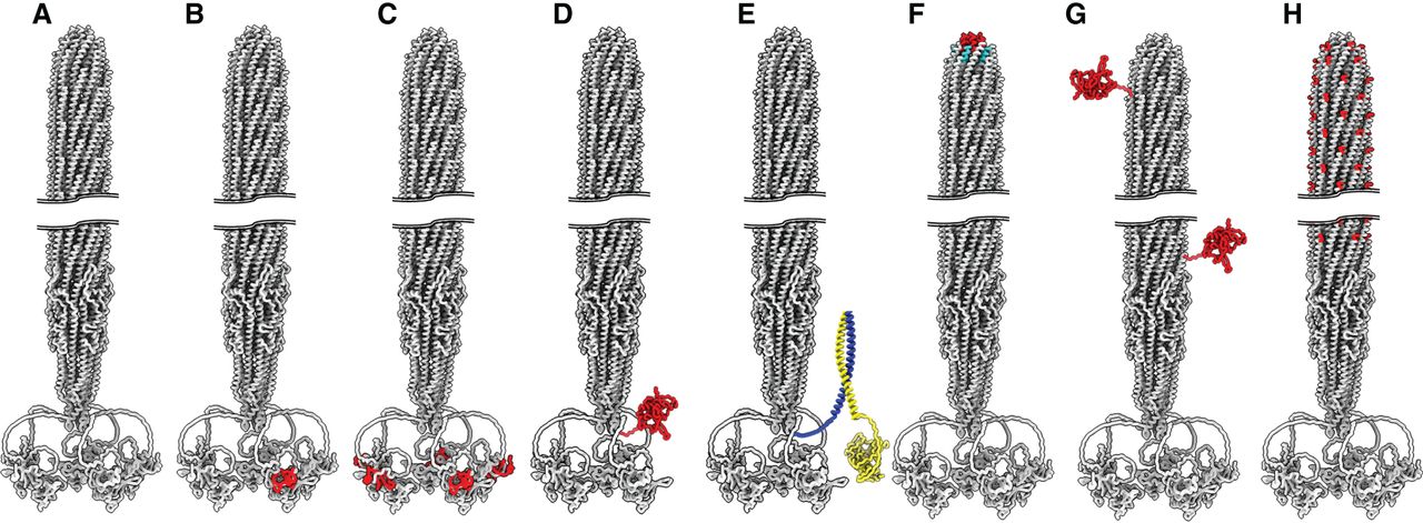
Positions of displayed proteins on the Ff virion. The wild-type Ff is shown in A and various sites and modes of display are shown in B–H. (B) Monovalent and (C) polyvalent display at the amino terminus of full-length pIII; (D) monovalent display on the C domain of pIII; (E) indirect phage display where the leucine zipper dimerization domain of Fos (blue) is fused to the pIII C domain, and the proteins to be displayed (yellow) are fused to the carboxyl terminus of its partner Jun (Jun and Fos Leu zipper coordinates are obtained from 5VPE [Yin et al. 2017]); (F) pVII (red) and /or pIX display (cyan); (G) mosaic pVIII display, where the fusions are assembled into the virion in combination with wild-type pVIII; (H) uniform pVIII display of short peptides on each pVIII subunit. The composite structure of an f1 phage virion was generated as described in Figure 1.


