
Cover image
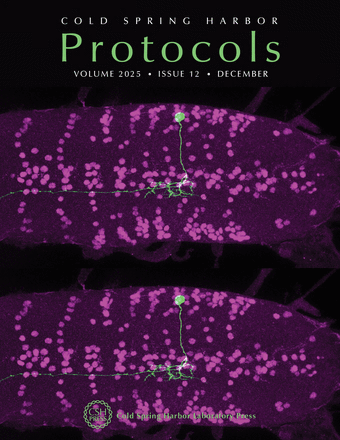
Nervous systems feature neurons with diverse shapes and complex spatial relationships, and the nervous system of Drosophila melanogaster provides a powerful model for characterizing neuronal diversity and connectivity. Visualizing the morphology of individual neurons is essential for understanding circuit architecture, yet it remains technically challenging due to the densely packed nature of neural tissue. In Drosophila, the multicolor flip-out (MCFO) technique allows the stochastic labeling of a limited number of neurons within a given GAL4 expression pattern, enabling single-cell resolution of neuronal morphology. In this issue, Marshall et al. describe a detailed protocol for performing MCFO in Drosophila larvae, from outlining the steps for crossing the MCFO driver fly line with the GAL4 line of interest, to image analysis (doi:10.1101/pdb.prot108422). The cover image shows a projection of a first instar Drosophila larval central nervous system stained with anti-Even-skipped (magenta) and anti-HA (green). An Even-skipped–positive lateral interneuron sends its neurite across the midline and terminates in the thoracic ventral nerve cord. Single-cell morphology was captured using the MCFO technique. This particular morphology is uniquely identified in the connectome as A08v. Image provided by the authors.

