
Cover image
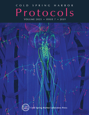
The Drosophila melanogaster larval neuromuscular junction (NMJ)—a synapse formed between motor neurons and skeletal muscle fibers—is a powerful system for investigating molecular mechanisms of synaptic growth and plasticity, and it is a great model to study normal development and neurological diseases. One of its key advantages is its relative simplicity: The muscles are large and arranged in an invariant, segmentally repeating pattern, and each muscle is innervated by the same identified motor neurons. Furthermore, the NMJs are individually specified, and easy to visualize and record from. Fundamental mechanisms underlying neuronal function have been uncovered at the fly's NMJ, and new pathways continue to be uncovered. These discoveries are fueled by the ease of dissections and an extensive array of techniques, including microscopy approaches. In this issue, Ashley and Carrillo describe how to use immunolabeling to visualize embryonic/larval presynaptic and postsynaptic structures at the Drosophila NMJ (doi:10.1101/pdb.prot108500). The cover image shows a Drosophila larval body wall and ventral nerve cord. UAS-GFP (green) is expressed by A8-GAL4, which is a fragment of the DIP-α promoter. A8-GAL4 drives expression in a subset of neurons, including the segmentally repeated phasic motor neurons that innervate dorsal or ventral muscle fields. The prep is also labeled with anti-DLG (red), a postsynaptic neuromuscular junction protein, and phalloidin (blue), to visualize F-actin and all muscles. Image provided by the authors.

