
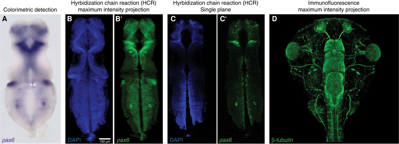
Figure 3.
Sample RNA in situ hybridization and immunostaining micrographs. (A) Colorimetric staining for pax6 RNA (purple) in a stage 46 Xenopus tropicalis dissected brain imaged by widefield microscopy. (B,C) Fluorescence staining for pax6 RNA by HCR (green; B′, C′) costained with DAPI to label nuclei (blue; B, C) in a stage 46 X. tropicalis dissected brain imaged by confocal microscopy. (B–B′) Maximum intensity projection of confocal sections. (C–C′) Single imaging plane. Note the increased resolution potential with the fluorescence-based method. (D) Immunostaining for β-tubulin in the stage 46 X. tropicalis head region imaged by confocal microscopy.


