
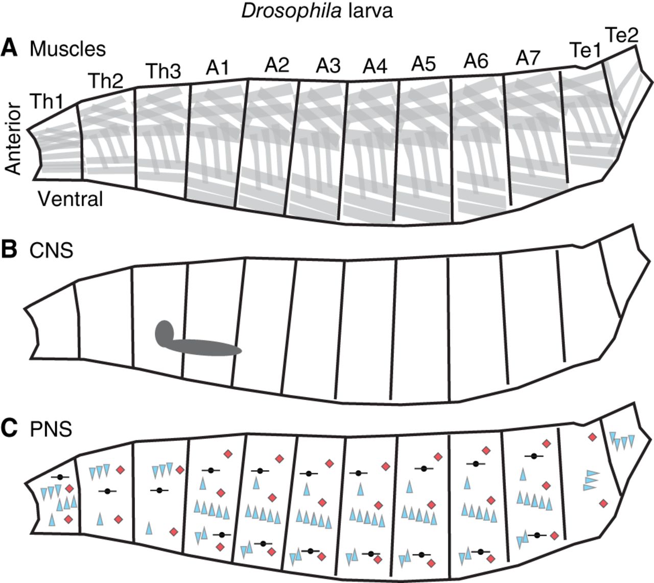
The organization of the Drosophila larval body. Illustrations of the Drosophila larval body are shown in the side view with anterior to the left and dorsal up. The body is left–right symmetrical and segmentally repeated. Segments are grouped into regions, and each segment within a region is numbered. Here, regions are Th (thorax), A (abdomen), and Te (terminus). (A) Organization of Drosophila muscles, with each gray box representing a muscle cell. Muscles are grouped into dorsal, lateral, and ventral sets, which repeat along the body axis. Not all muscles are shown. (B) The illustration shows the position of the larval central nervous system (CNS in gray), with brain lobes (left, circular) and nerve cord (right, elongated circle). (C) The illustration shows the larval peripheral nervous system (PNS) with different classes of primary somatosensory neurons displayed as symbols. Chordotonal neurons (vibration sensors) are shown as blue triangles. Red diamonds represent multidendritic class four neurons (nociceptors). Dorsal bipolar dendrite neurons (stretch receptors) are shown in black. The distribution of primary somatosensory neurons is identical in abdominal segments but diverges in the thorax and terminus. Not all sensory neuron types are shown.


