
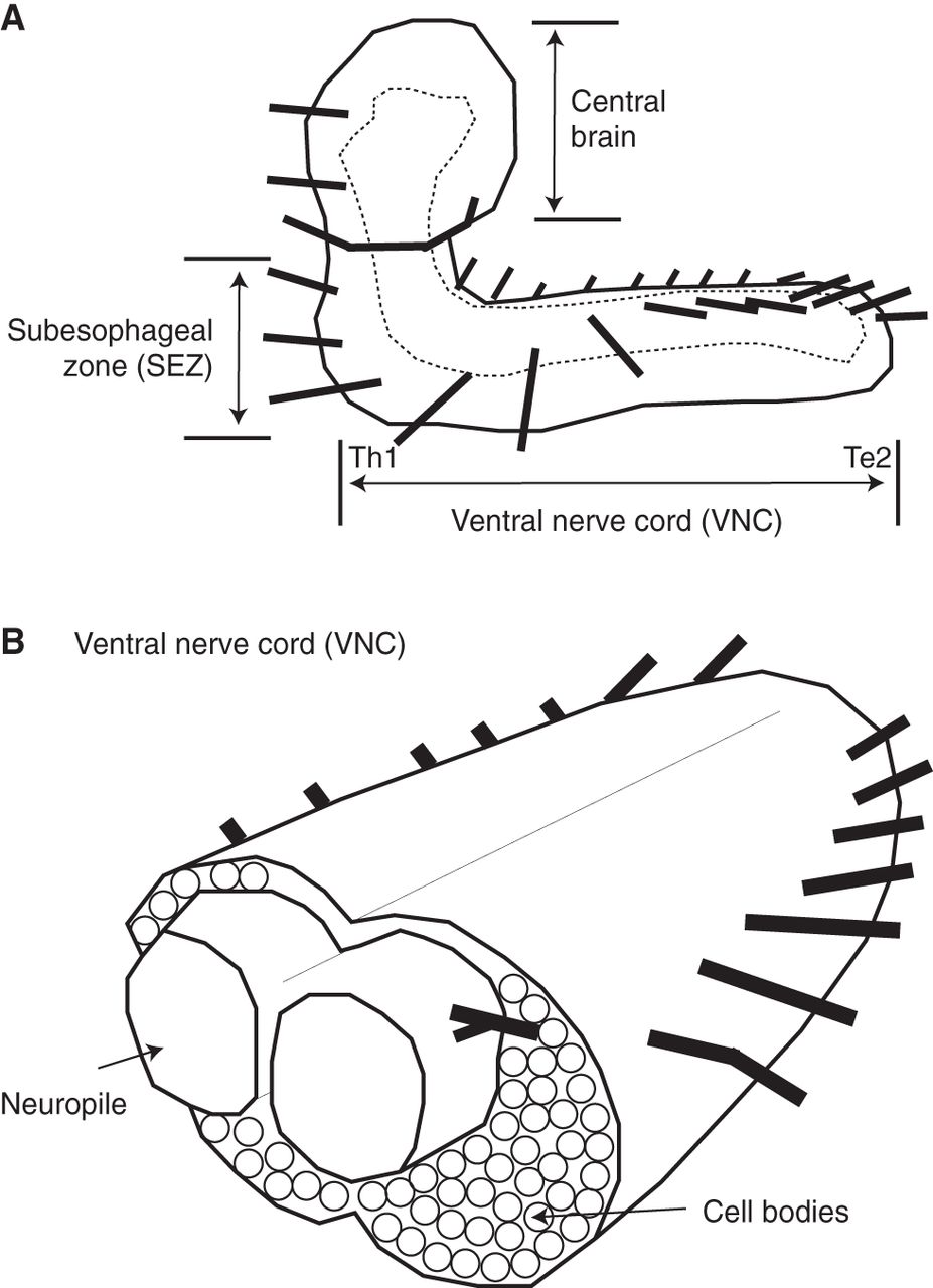
The anatomy of the Drosophila larval central nervous system (CNS). (A) An illustration of the Drosophila larval CNS is shown in a side view, anterior to the left and dorsal to the top. The dashed line represents the border of the neuropile. Each black line projecting from the CNS represents a nerve root. There is one nerve root per segment. The three major CNS regions are labeled (central brain, SEZ [subesophageal zone], VNC [ventral nerve cord]). Motor circuits are housed in the ventral nerve cord. (B) A diagram of a cross-section of the VNC is shown, anterior to the left. The neuropile is an axon-, dendrite-, and synapse-rich region. Cell bodies are peripheral to the neuropile. Each black line projecting from the CNS represents a nerve root.


